Naturally occurring melanomas in dogs as models for non-UV pathways of human melanomas
Marc Gillard 1,2, Edouard Cadieu 1,2, Clotilde De Brito 1,2,
Jérôme Abadie 3, Béatrice Vergier 4, Anne-Sophie Guillory
1,2, Patrick Devauchelle 5, Frédérique Degorce 6,
Laëtitia Lagoutte 1,2, Benoit Hédan 1,2, Marie-Dominique Galibert
1,2, Francis Gallibert 1,2 et Catherine André 1,2
1 CNRS, UMR 6290, Institut de Génétique et Développement de Rennes, Rennes,
France.
2 Université Rennes 1, UEB, IFR140, Faculté de Médecine, Rennes, France.
3 Laboratoire d’Histopathologie Animale, Oniris, Ecole Nationale Vétérinaire,
Nantes, France.
4 Service de Pathologie, CHU Bordeaux and Université Bordeaux Segalen,
France.
5 Centre de Cancérologie Vétérinaire, ENVA, Maisons Alfort, France.
6 Laboratoire d’Anatomie Pathologique Vétérinaire du Sud-Ouest LAPVSO,
Toulouse, France.
Summary
Spontaneously occurring melanomas are frequent in dogs. They appear at the same
localizations as in humans, i.e. skin, mucosal sites, nail matrix and eyes. They
display variable behaviors: tumors at oral localizations are more frequent and aggressive
than at other anatomical sites. Interestingly, dog melanomas are associated with
strong breed predispositions and overrepresentation of black-coated dogs. Epidemiological
analysis of 2350 affected dogs showed that poodles are at high risk of developing
oral melanoma, while schnauzers or Beauce shepherds mostly developped cutaneous
melanoma. Clinical and histopathological analyses were performed on a cohort of
153 cases with a 4-yr follow-up. Histopathological characterization showed that
most canine tumors are intradermal and homologous to human rare morphological melanomas
types – ‘nevocytoid type’ and ‘animal type’-. Tumor cDNA sequencing data, obtained
from 95 dogs for six genes, relevant to human melanoma classification, detected
somatic mutations in oral melanoma, in NRAS and PTEN genes, at human hotspot sites,
but not in BRAF. Altogether, these findings support the relevance of the dog model
for comparative oncology of melanomas, especially for the elucidation of non-UV
induced pathways.
Review
The first original article on this subject, this study comparing melanomas in dogs
and humans underlines the value of comparative oncology for both human and veterinary
medicine. The article reveals that certain dog breeds are more likely to develop
certain types of melanomas, while other breeds only very rarely suffer from them.
This finding could result in vets and dog owners becoming more aware of the possibility
of melanomas occurring in certain breeds. It should also allow researchers to identify
genetic alterations specific to certain breeds likely to develop melanomas. A research
programme into the genetics of melanomas in dogs has been running at the CNRS in
Rennes for several years, with a team of vets, doctors, experts in veterinary and
human anatomopathology and geneticists working on it. This work was initiated at
the CNRS in Rennes under the leadership of Dr Catherine André and in collaboration
with Dr Patrick Devauchelle (MICEN-Vet, Créteil, France), Dr Jérôme Abadie (Nantes
Veterinary School, Oniris, France) and with the help of the French veterinary network
AFVAC (www.AFVAC.com) and the Cani-DNA biobank (http://dog-genetics.genouest.org)
developed at the CNRS in Rennes. To date, this research has resulted in a PhD thesis
in 2011, several papers presented at veterinary and human genetics congresses and,
most recently, the publication of the scientific article we are summarising here.
Apart from the genetic analyses, a further research objective is to find ways of
better treating melanomas, both in dogs and in humans. Numerous types of melanomas
are to be found in both species, whereby in dogs the most frequent and most severe
ones are oral melanomas. Such melanomas lend themselves to genetic and therapeutic
studies in dogs and humans for whom melanoma tumours located in the mouth are rare
and poorly documented at the genetic level. The majority of cutaneous melanomas
in dogs are intra-dermal and of a dark colour. This study of dogs helps us better
understand genetic causes of melanomas not linked to exposure to the sun and potentially
to develop new therapies benefiting both dogs and humans.
Running for more than 5 years, the current research programme in the Rennes laboratory
has been financed by the CNRS, the Brittany region (a PhD grant), the French Anti-Cancer
League (post-doctoral funding) and the NCIa, the French National Cancer Institute
which has funded two research projects. Also involved are numerous vets, breeders
and dog-owners.
This issue of Pigment Cell and Melanoma Research, published in January 2014,
focuses on the value of comparative studies of melanomas in dogs and humans, witnessed
by the journal's cover created at the Rennes laboratory (Cadieu et al.), an editorial
and a "perspective" on the same subject.
The article is available on
http://onlinelibrary.wiley.com/doi/10.1111/pcmr.12170/abstract
Gillard et al. 2013-PCMR Cover
© Photos credits to (upper left to upper right): Dr T. Jouary (1, 3); Dr P. Devauchelle (2); Dr M. Delverdier (4, 10); Pr A. Dupuy (5, 7, 9); Pr F. Fogel (6); Dr A. Muller (8); I. Raymond (11).
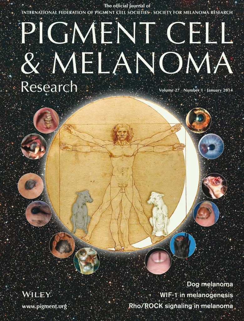

The dark side of human melanoma. The Vitruvian Man and dogs, drawn by Leonardo da Vinci, accent the power of the dog model to shed light on non-UV light dependent human melanoma. The additional images illustrate the striking correspondences between human and dog anatomic sites: mucosal/oral; cutaneous; acral (soles/footpad); nail matrix; acral (digit); eye. Image composition is from Cadieu et al.
To participate in this research by sending in samples and/or information:
Contact : Edouard Cadieu, Clotilde de Brito
Tel +33 2 23 23 45 09 Fax : +33 2 23 23 44 78
Website on "dog genetics":
http://dog-genetics.genouest.org